Techniques
The Manns Lab focuses on the study of memory. We use a number of techniques to pursue our research questions.
Memory testing in rats
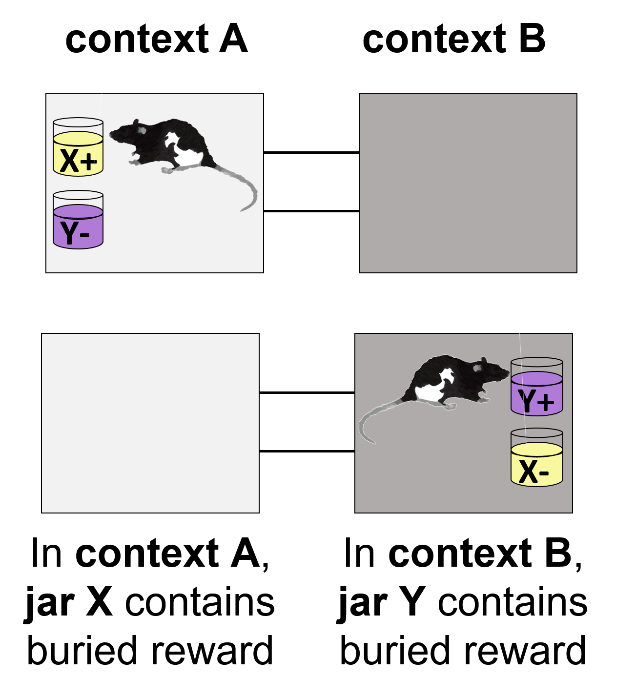
We have used a variety of tasks to assess memory in rats. Examples have included novel object recognition memory, object-location associative memory, object-context associative memory (pictured), and memory for temporal order of odors. We use a variety of sophisticated techniques to record and perturb brain activity, but testing memory in rats is easily our most challenging technique.
Memory testing in humans
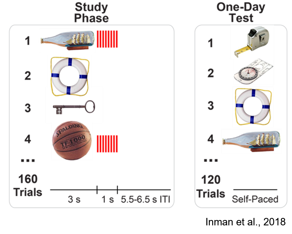
We have used a variety of tasks to assess memory in humans. Examples have included novel object recognition memory (pictured) and semantic memory for factual knowledge.
Tetrode-based single-unit electrophysiology
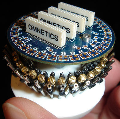
We use custom-built driver assemblies (pictured) to be able to position up to 32 tetrodes independently in target rat brain regions. The advantage of tetrodes (a bundle of four electrodes) is that they enable sorting of action potentials into separate clusters thought to represent individual neurons (called units in the jargon of the field). Accordingly, one can ask questions about how ensembles of neurons represent memories.
Silicon probes
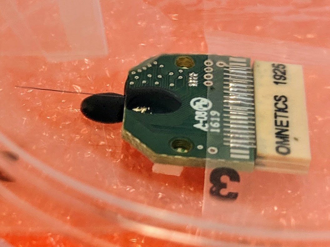
We also use silicon probes to record electrical activity from brains as rats perform memory tasks. The advantage of silicon probes is that they allow for a densely-packed and regularly-spaced array of electrode contacts along the shank of the probe. As a result, one can ask questions about, for example, layer-specific oscillations in the hippocampus. We are currently using both low-cost 32-channel probes as well as more expensive 1000-channel probes (Neuropixels), depending on the experiment.
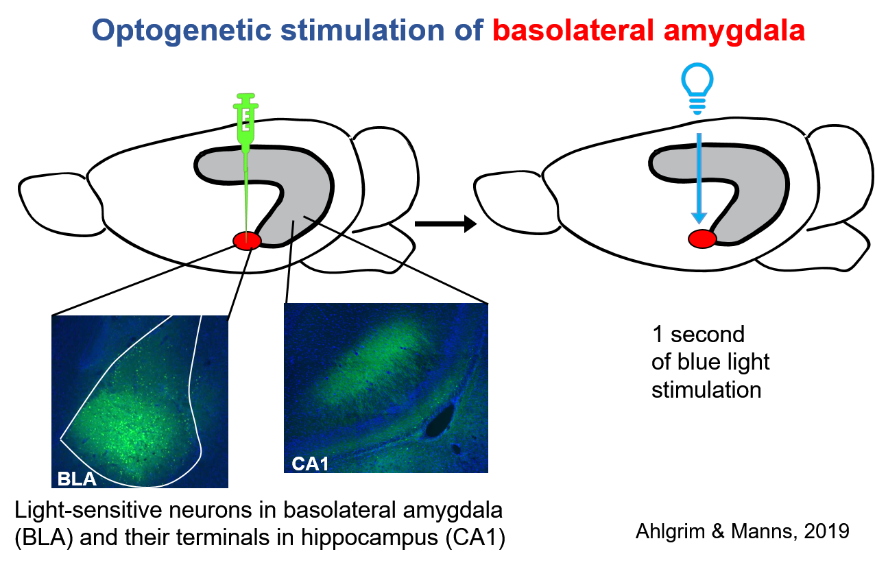
Optogenetic brain stimulation
Optogenetics involves transfecting specific neurons with genetic material that eventually leads to those neurons being sensitive to light. Thus, one can stimulate specific neurons such as glutamatergic projection neurons in the basolateral amygdala. We have used optogenetics in combination with electrophysiological recordings to ask how stimulation of the basolateral amygdala modulates memory-related activity in the hippocampus.
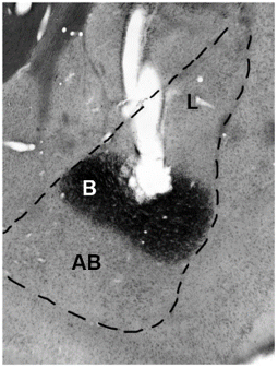
Electrical brain stimulation
We have used electrical brain stimulation to deliver brief (1 second) and very low-level current (20 microamps in rats; 500 microamps in humans) to the basolateral amygdala in both rats and humans to enhance object recognition memory. Indeed, the advantage of electrical stimulation over optogenetic stimulation is that electrical stimulation can be used similarly in rats and humans. The image depicts the postmortem marking lesion made by a bipolar stimulating electrode implanted in a rat's basolateral complex of the amygdala (BLA), which is outlined with a dashed line. The tissue was processed with a stain (acetylcholinesterase stain), which helps distinguish the nuclei (B: basal; L: lateral; AB: accessory basal) in the BLA.
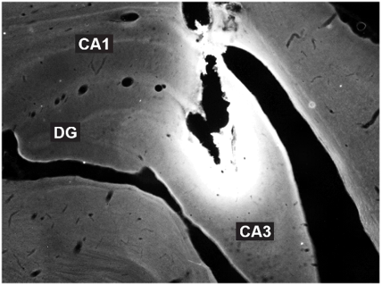
Local brain infusions
We have used chronically-implanted cannula to be able to deliver drugs directly to specific brain regions. The advantage of this approach is that it allows one to induce reversible brain changes and compare the effects to a control condition in which only saline is infused into the brain region. For example, we have infused muscimol to temporarily inactivate the hippocampus to ask about its role in amygdala-mediated memory enhancement. The image depicts a section of the hippocampus imaged under a fluorescent microscope, which shows the spread of the fluorophore-conjugated muscimol.
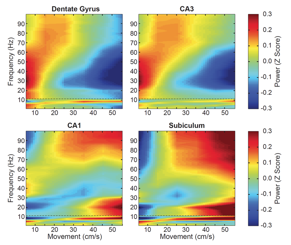
Analyses of neural data
For some experiments, the work is only half done after the data are collected. Our lab uses a variety of analytical approaches to analyze behavior and brain signals, including spiking activity and local field potentials. The image to the left shows the relationship between oscillatory power and rat locomotive speed in four hippocampal regions.